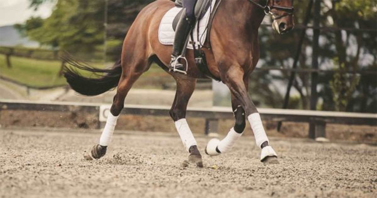
Navicular disease (ND) is a chronic degenerative condition of the navicular bone of the foot and is one of the most common and frustrating causes of horse lameness. The syndrome appears to have complex pathogenesis (cause) rather than being a specific disease entity. The consensus is that whilst a significant component is biomechanical, there is also a vascular component as well.
It is generally considered a significant joint problem in the more mature riding horse, commonly appearing around 8 to 12 years of age. There is also an hereditary component as navicular disease is overrepresented in some breeds (e.g. Warmblood horses, Quarter horses, and Thoroughbreds) and rare in others (e.g. Arabs, Friesians). It is usually a progressively deteriorating condition.
Conformation of the distal limb is likely to play a large and significant role in the process and the degree of lameness. Excessive pressure on the navicular bone occurs with a “broken back” hoof-pastern axis, usually accompanied by an underrun heel and excessively long toe. This conformation leads to excessive concussion between the flexor tendon and the navicular bone. It may also cause navicular bursitis, with direct damage to the flexor surface’s fibrocartilage and the collagenous surface of the flexor tendon itself.
How to tell if your horse has Navicular Disease
The disease is usually gradual in onset. Owners typically report an intermittent, shifting lameness and a reluctance to work. Because the disease is commonly bilateral, there may be no obvious head nod when the horse is trotted in a straight line, with only a shortened stride present. Horses typically tighten through the neck muscles and become stiff and may even change their behaviour due to being painful and uncomfortable in both front limbs.
Lameness is usually exacerbated by lunging the horse in a circle, which can be a useful aid to diagnosis. In the early stages of the disease, the lameness may not be visible even at a lunge and until a nerve block is performed on one limb (i.e. the two lame feet cancel each other out). A flexion test of the distal forelimb may produce a transient worsening of the lameness but can be quite subjective.
Clinical Diagnosis
Clinical diagnosis by a veterinarian is crucial. This is mainly based on the horse’s presentation (age, breed commonly at risk) and, significantly, on the lameness examination.
Horses with navicular disease are rarely positive to hoof testers, but a palmar digital nerve block will eliminate the lameness.
This nerve block anesthetises the entire sole and coffin joint in addition to the heel, so a response to the block itself is not diagnostic. A transfer of lameness to the other forelimb, which is also eliminated by a palmar digital nerve block, is necessary for a tentative diagnosis of navicular disease. Anaesthesia of the navicular bursa is much more specific but not as commonly performed because of the level of difficulty with this procedure.
Radiographs should be taken but do not always correlate directly to the severity of lameness or disease. Radiographs can demonstrate a range of degenerative joint changes in the navicular bone: marginal enthesiophytes, enlarged synovial fossae (so-called vascular channels) of variable size and cysts due to loss of medullary trabecular bone, general sclerosis of the medullary cavity, and flexor surface changes (observed on “skyline” radiographic views), including erosions and loss of a defined cortex.
Advanced imaging (Magnetic Resonance Imaging and Computed Tomography) can offer superior diagnostics for soft tissue injuries inside the hoof capsule. These appear particularly useful for picking up subtle lesions earlier in the disease process and that can impact prognosis. These diagnostics are not always readily available or economically viable for some owners.
What is the best treatment for navicular disease in horses
It is important to remember that navicular disease is both chronic and degenerative, so although it can be managed in some horses, it cannot be cured.
Corrective shoeing is paramount and getting your farrier and vet to work together is essential. The concept is trimming and shoeing to restore normal alignment in the foot (hoof-pastern axis) and decrease pressure on the navicular area. This typically takes a number (up to 3), shoeing’s to achieve. The shoeing technique that most effectively reduces pressure on the navicular area is raising the heel (usually performed with wedge pads or a wedged shoe). Rolling the toe of the shoe further relieves the pressure on the navicular bone and improves break over. The egg bar shoe does not decrease navicular pressure in sound horses on a hard surface. Still, it can effectively decrease the navicular bone forces in some horses with navicular disease or collapsed heels. Additionally, egg bar shoes are more likely to effectively decrease forces on the navicular area on soft surfaces (that horses are often worked on). That said, egg bars can be difficult to maintain and hold on so sometimes a straight bar shoe may also be considered adequate in some cases.
Non-steroidal anti-inflammatories (NSAID’s) are used during this period to manage the pain and help get the horse moving which may also help with vascular flow in the region. Strict rest is not recommended (unless severe lameness is present or persists) and a controlled and graduated exercise programme essential to the horses rehabilitation.
Intra-articular medications with corticosteroids can also be useful. These are most commonly directed into the coffin joint (reported 33% successful for 2 months). In contrast, injection directly into the navicular bursa itself has a better response (80% successful for up to 4 months), but as mentioned before, is technically more difficult and usually performed under radiographic guidance. Recently, Arthramid 2.5% iPAAG used intraarticularly has also been reported for the treatment for navicular disease with excellent results. Dr Andy Bathe, Rossdale and Partners, UK presented three cases non-responsive to corticosteroids that had good long-term results; Veterinary and Comparative Orthopaedics and Traumatology Abstract.
Surgical options such as digital neurectomy are available but carry numerous risks and are considered ethically unjustifiable by many. This procedure excludes the horse from FEI or racing competitions.
Summary
So although the prognosis is considered guarded to poor for horses with navicular disease, a carefully designed therapeutic regimen, including corrective shoeing, NSAID therapy, and appropriate navicular bursa injections (and/or possibly coffin joint injections), can prolong the usefulness of most horses and the competitive status of many. Over months or years, most affected horses though will reach a point of non-responsiveness to treatment.


