In this second issue we report on how an experienced racetrack veterinarian managed a case of chronic tenosynovitis of the deep digital flexor tendon sheath in a racing Thoroughbred.
Case History:
An 8 year-old TB racehorse presented with subacute- chronic digital flexor tendon sheath effusion in the right forelimb. The horse had been on and off lame for about 4 months, had mild pain on flexion, a shifting gait at canter and gallop and was reluctant to lead on the affected limb.
The effusion was made worse after fast work, and although it improved temporarily to treatment with triamcinolone/ HA (for 3-4 weeks), it worsened again with increasing galloping exercise later in its preparation.
Diagnostic Investigation:
Radiography and ultrasonography of the fetlock and surrounding structures was performed and detected no abnormal findings (apart from the tenosynovitis). Further diagnostics were offered (MRI, tenoscopy) but declined by the owner’s agent.
Treatment:
The area was prepped aseptically and 2mls (50mg) of 2.5% iPAAG Arthramid was injected directly into the tendon sheath. This was followed by 3 days of swimming exercise before the horse returned to full work (galloping exercise).
Outcome:
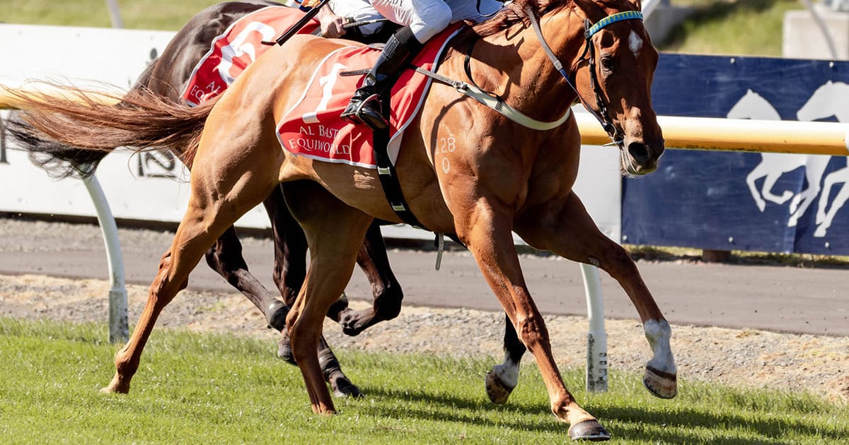
Re-examination after 18 days revealed a significant reduction in the degree of tenosynovitis and the horse was no longer reactive to flexion. The track rider reported the horse to be working evenly and would now lead consistently on the affected limb. A prolonged response to treatment was seen under full race training and the horse was racing and winning.
Discussion:
There have been no case reports to date on the clinical application of 2.5% iPAAG Arthramid for managing tenosynovitis of the digital flexor tendon sheath.
Ultrasonography is an excellent tool for imaging this area but has relatively low sensitivity for detecting longitudinal tears in the tendon or paratenon of the deep digital flexor tendon itself. This could have been missed and would have carried a worse prognosis without tenoscopy.
Most likely this was a case of primary synovitis of the tendon sheath itself that became chronic over time. This may occur due to poor conformation in conjunction with heavy workloads, or acute injuries that tend to not resolve due to ongoing inflammation of the synovial lining of the tendon sheath.
Wehling et al. (2007) showed mononuclear cells respond to a pyrogenic-free substance, such as 2.5% iPAAG, with the release of anti-inflammatory cytokines, such as interleukin¹ receptor antagonist protein, transforming growth factor¹, and insulin-like growth factor¹, amongst many other cytokines1. This may explain the reduction in tenosynovitis seen in this case. It is also known that 2.5% iPAAG acts as a dynamic tissue scaffold, integrating into tissues themselves by a combination of cell migration and vessel in growth, that in turn increases tissue elastance². In articular joints, it is further documented that a new and hypercellular synovial cell lining forms on the internal surface. Therefore, it is possible that administration of 2.5% iPAAG Arthramid in this case resulted in improved function of the tendon sheath and an improvement in the quality of synovial fluid leading to a resolution of the synovitis. Further studies are necessary and should include quantitative assessment of the quality of synovial fluid.
Dr Jason Lowe BVsc, Cert EP, MBA.


